A BANANA THROUGHT THE LENS OF MEDICAL IMAGING
This creative visual shows how different diagnostic tools visualize the same object:
Top Left: The banana in normal visible light
– what our eyes see. 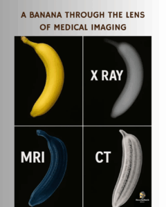
Top Right (X-RAY): Captures dense structures; mostly shows the banana’s outer shape.
Bottom Left (MRI): Reveals soft tissue details using magnetic fields – you can see the internal texture and fibrous structure inside the banana.
Bottom Right (CT): Uses X-rays to create detailed cross-sections – displays inner density variations and even tiny internal differences in pulp distribution.
Perfect for understanding how each scan works in real diagnostics!
Source: world_of_biology_wob